Upper Wisdom Teeth and Your Sinuses: What Patients Should Know
Upper wisdom teeth sit remarkably close to your sinuses. Learn what that means for your surgery, what a sinus communication is, and why it's usually far less serious than it sounds.
Anatomy
Most patients researching wisdom teeth removal focus on the lower teeth — the nerve proximity, the higher dry socket risk, the more complex impactions. Upper wisdom teeth get comparatively little attention, which means one of their defining anatomical features often comes as a surprise: they sit remarkably close to your maxillary sinuses.
This article explains that relationship in plain terms, what it means for your surgery, what happens if a sinus communication occurs, and why it is usually far less alarming than the name suggests.
Key Takeaways
The maxillary sinuses sit directly above the upper back teeth — in some patients, the roots of upper wisdom teeth extend into or against the sinus floor.
A small sinus communication during upper wisdom tooth extraction is a recognized, manageable occurrence — not a surgical error.
Most small communications close on their own within a few weeks with proper precautions.
Sinus precautions after upper extractions — no nose blowing, no straws, sneeze with your mouth open — protect the healing site.
A panoramic X-ray, and sometimes a CBCT scan, allows the surgical team to anticipate sinus proximity before the procedure.
The Anatomy: Why Upper Wisdom Teeth Are Different
Where the Maxillary Sinuses Sit
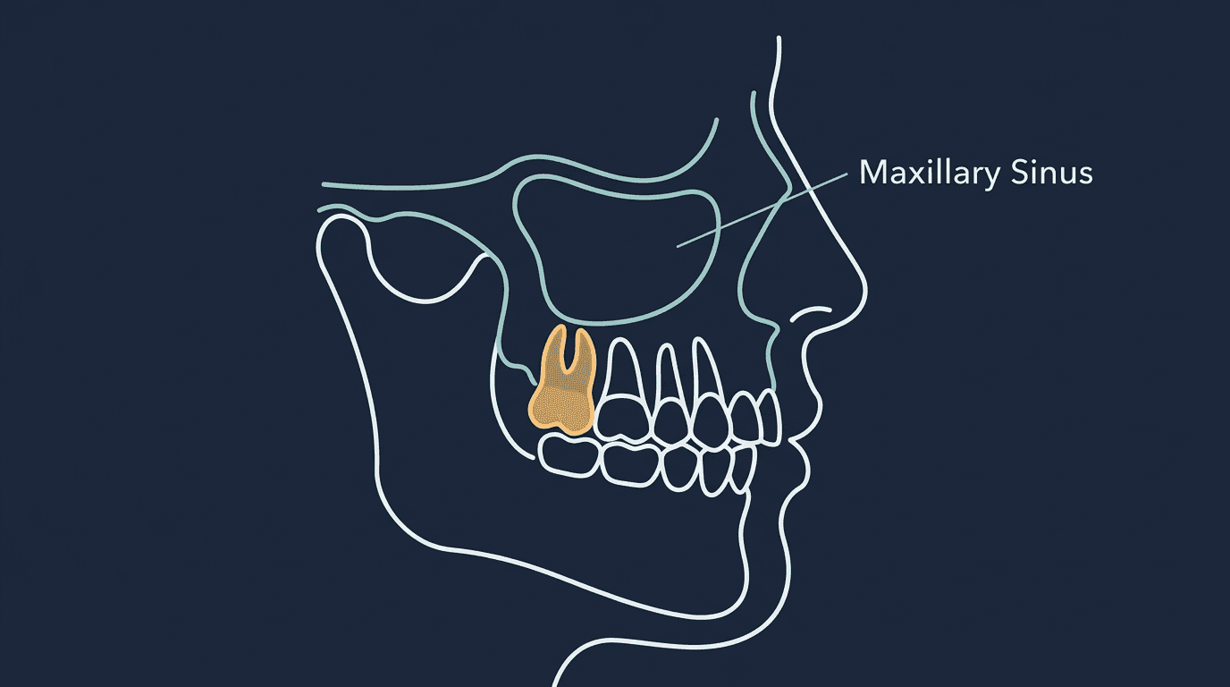
The maxillary sinuses are two air-filled cavities located within the cheekbones, one on each side, directly above the upper back teeth. They are the largest of the paranasal sinuses and serve functions including humidifying inhaled air, lightening the weight of the skull, and contributing to voice resonance.
The floor of each maxillary sinus forms a thin bony boundary above the roots of the upper molars — including the upper wisdom teeth. In many patients, that boundary is only a millimeter or two thick. In some, the tooth roots actually protrude into the sinus space, separated from the sinus lining by nothing more than a thin membrane.
How This Varies Between Patients
Sinus anatomy differs considerably from person to person. Some patients have small sinuses well separated from their tooth roots. Others have large, pneumatized sinuses extending down between the roots of the upper molars. This variation is one reason a current panoramic X-ray matters before any upper wisdom tooth extraction — it shows the specific relationship in your case rather than relying on a general assumption.
For cases where the relationship appears particularly close on a panoramic image, a CBCT (cone beam computed tomography) scan may be ordered. This provides three-dimensional detail that a standard two-dimensional panoramic X-ray cannot, allowing more precise surgical planning.
What Is an Oroantral Communication?
An oroantral communication is a small opening between the mouth and the maxillary sinus that can occur when an upper tooth with roots close to the sinus is removed. When the tooth comes out, the thin bone and membrane separating the socket from the sinus can be disrupted, leaving a small passage between the two spaces.
The clinical terminology sounds more alarming than the reality for most cases. Small communications are a recognized possible outcome of upper molar extraction — particularly for wisdom teeth — rather than a sign that something went wrong. Surgeons anticipate this possibility, check for it during the procedure, and manage it when it occurs.
How It's Identified
Surgeons check for a communication at the time of extraction using a simple, non-invasive assessment. If one is present, it is identified immediately — not discovered later — which allows appropriate management from the start.
How Small Communications Are Managed
The management depends on size:
Very small communications: Often require no active intervention beyond protecting the blood clot and following sinus precautions. The site typically closes on its own within a few weeks as normal healing progresses.
Moderate communications: May be managed with additional closure measures at the time of surgery — such as suturing the gum tissue to fully cover the socket, or placing material in the socket to support closure. PRF, which is placed in every socket at Dr. Wisdom Teeth, contributes to this healing process.
Larger communications: Less common with wisdom teeth specifically, and may require a more involved closure procedure. These are the exception rather than the norm.

Sinus Precautions: What to Follow After Upper Wisdom Tooth Removal
If a sinus communication occurred — or if your surgeon determined that your sinus anatomy makes one possible — you'll be given specific precautions. These are designed to prevent pressure changes between the mouth and sinus that could disrupt healing.
No forceful nose blowing. Typically for about two weeks. Blowing your nose creates pressure that can push through a healing communication. Gently wipe instead.
Sneeze with your mouth open. This releases pressure through the mouth rather than driving it into the sinus and the healing site.
No straws, no smoking, no suction. The same rule that protects against dry socket also protects a healing sinus communication.
Avoid heavy lifting and straining. Activities that significantly increase pressure — heavy weightlifting, straining — should be paused for the period your surgeon specifies.
Discuss air travel and diving. Both involve significant pressure changes. If you have travel planned shortly after surgery, mention it to your surgical team so they can advise on timing.
Take prescribed medications as directed. This may include a decongestant or antibiotic depending on your specific situation.
Have upper wisdom teeth and questions about your sinus anatomy? A consultation with current imaging gives you a clear picture. Call (801) 370-0050 or book at drwisdomteeth.com.
Sinus-Related Symptoms After Upper Extractions
Some sinus-adjacent sensations after upper wisdom tooth removal are expected and resolve as part of normal healing. Others warrant a call to your surgical team.
Generally Normal
Mild pressure or fullness in the cheek area for a few days
Slight congestion on the side where extraction occurred
A small amount of blood-tinged nasal discharge in the first day or two
Mild sensitivity when bending forward
Contact Your Surgical Team If You Experience
Air or liquid passing from your mouth into your nose, or vice versa — for example, water coming out of your nose when drinking
A persistent salty or unusual taste that doesn't resolve
Increasing facial pressure or pain after day 3 rather than improving
Thick, foul-smelling nasal discharge, which can indicate a sinus infection
Fever accompanying sinus symptoms
A whistling sound when speaking or breathing through the extraction area
These signs don't necessarily indicate a serious problem, but they do warrant evaluation. Sinus complications after extraction are highly treatable when addressed promptly.
Can Wisdom Teeth Cause Sinus Problems Before Removal?
Yes — the relationship works in both directions. Infected or impacted upper wisdom teeth can contribute to sinus symptoms, a condition sometimes described as sinusitis of dental origin.
Because the tooth roots sit so close to the sinus floor, infection at the root of an upper wisdom tooth can extend into the sinus cavity. This produces symptoms that closely resemble ordinary sinusitis — facial pressure, congestion, discharge — but that don't respond fully to standard sinus treatment because the underlying source is dental.
Patients who experience recurrent one-sided sinus symptoms that don't fully resolve with typical treatment, particularly if they still have upper wisdom teeth, may benefit from a dental evaluation alongside the sinus workup. Addressing the dental source, when that's what it is, resolves symptoms that otherwise persist.
How Surgical Planning Accounts for Sinus Proximity
Sinus proximity is anticipated, not discovered mid-procedure. The planning process includes:
Panoramic X-ray review: Shows the general relationship between upper wisdom tooth roots and the sinus floor.
CBCT imaging when indicated: Three-dimensional imaging for cases where the panoramic image suggests particularly close proximity or unusual root anatomy.
Surgical technique selection: Approach and instrumentation are adapted based on the anticipated anatomy — including sectioning the tooth rather than applying force that could displace a root toward the sinus.
Intraoperative assessment: Checking the socket after extraction so any communication is identified and managed immediately.
PRF placement: Included in every socket at Dr. Wisdom Teeth, supporting clot stability and tissue healing — relevant to sinus-adjacent sites as well as lower extraction sockets.
Frequently Asked Questions
Does a sinus communication mean something went wrong during my surgery?
No. Small communications are a recognized possible outcome of upper molar extraction when tooth roots sit close to the sinus floor — an anatomical reality, not a technique failure. Surgeons anticipate this possibility, check for it, and manage it when it occurs.
How long does a sinus communication take to heal?
Small communications typically close on their own within a few weeks when sinus precautions are followed. Larger ones may take longer or require additional closure measures. Your surgical team will advise on the expected timeline for your specific case and may schedule a follow-up to confirm closure.
Can I fly after upper wisdom teeth removal?
Discuss this specifically with your surgical team, particularly if a sinus communication occurred. Cabin pressure changes affect sinus pressure, and flying too soon after an upper extraction with sinus involvement can disrupt healing. If you have travel booked, bring it up before your procedure so it can be factored into planning.
Are lower wisdom teeth safer than upper ones because of this?
Neither is inherently 'safer' — they simply carry different anatomical considerations. Upper wisdom teeth involve sinus proximity. Lower wisdom teeth involve proximity to the inferior alveolar nerve and a notably higher dry socket rate. Both are routine procedures with well-established management approaches when properly planned.
Will I need a CBCT scan for my upper wisdom teeth?
Not usually. A panoramic X-ray is sufficient for most cases. CBCT is ordered selectively when the panoramic image suggests unusually close sinus proximity, unusual root anatomy, or another factor warranting three-dimensional assessment before surgery.
The Bottom Line
The close relationship between upper wisdom teeth and the maxillary sinuses is a normal anatomical fact, not a hidden danger. It's accounted for in surgical planning through imaging, technique selection, and intraoperative assessment. When a small communication does occur, it's identified immediately and managed with straightforward measures — most close on their own within weeks.
What matters practically is following the sinus precautions you're given: no forceful nose blowing, no straws, sneeze with your mouth open, and avoid straining. And if you notice air or liquid passing between your mouth and nose, or worsening facial pressure after day 3, call rather than waiting it out.
Schedule your consultation with current imaging: drwisdomteeth.com | (801) 370-0050 | Mon–Fri 8am–5pmProvo: 2230 N University Pkwy #8A | Murray: 5888 S 900 E #101

Written by
Dr. Wisdom Teeth
Resources

